
Innovative Techniques in the CoE
During its 300-year history, microbiology’s level of understanding has always reflected the availability of suitable methods for studying the structure and function of highly complex communities of organisms that are mostly invisible to the human eye.
Many of the CoE members are particularly strong in developing innovative methods for microbiome research, and the research in this cluster will be supported by nine dedicated non-profit facilities that form our cross-cutting methods module.
The facilities will boost research activities in all three themes, guaranteeing that comparable high-end data on microbiome structure and function will be gained within individual projects. All facilities will also offer education and training opportunities for early career researchers as well as workshops where users can share results and experiences.
MF1 Joint Microbiome Facility
State-of-the-art DNA/RNA sequencing
The JMF, coordinated by David Berry and Petra Pjevac, offers individualized consulting and services in study design, sample processing, sequencing, bioinformatic analyses, and data interpretation to both clinical and environmental microbiome researchers.
The JMF offers DNA/RNA extraction from diverse sample types, amplicon sequencing, full-length primer-free rRNA profiling, and metagenomic and metatranscriptomic sequencing using Illumina and Oxford Nanopore technologies.
The JMF is also developing and implementing amplicon approaches that allow for absolute, fully integrated ultra-high throughput long-read sequencing and activity-based sequencing approaches based on the incorporation of bioorthogonal or isotope-labeled nucleotides.
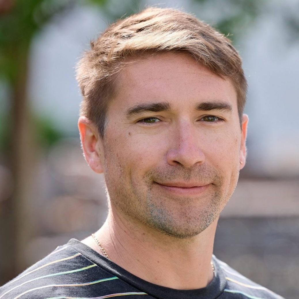
david berry
MF1 Coordinator
university of vienna
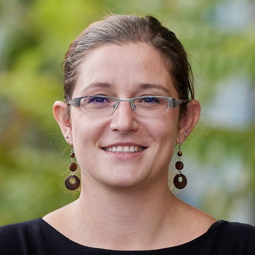
petra pjevac
MF1 Coordinator
university of vienna

maria de los Angeles Tarazona Montoya
Technical Assistant MF1
UNIVERSITY of Vienna
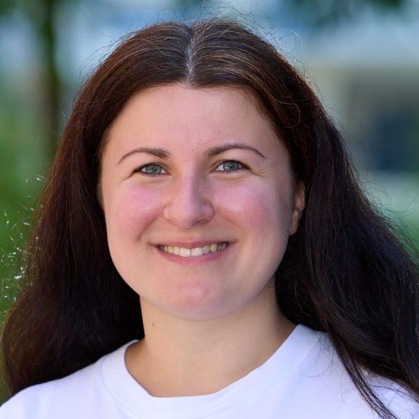
sara malinowski
Technical Assistant MF1
university of vienna
MF2 Bioanalytics and Environmental Mass Spectrometry Facility
State-of-the-art support in metabolomics and trace elements analysis
The facility consists of two subunits, the Bioanalytics, and the Environmental Mass Spectrometry units covering omics-scale biomolecular analyses and trace molecular and elemental analyses, respectively.
In the Bioanalytics unit at TU Wien, we perform proteomics, lipidomics, and metabolomics employing liquid chromatography ((nano-)UPLC) coupled to trapped ion mobility-tandem mass spectrometry on time-of-flight analyzers (timsTOF pro/HT). The Bioanalytics unit has a special focus on activity-based proteomics and multi-omics method development including sophisticated sample preparation, measurement methods, and data analysis workflows.
The Environmental unit at the University of Vienna performs environmental trace analysis and also covers the full analytical cycle. They currently run gas- and liquid chromatographic separation coupled to triple quadrupole, tandem mass spectrometry, inductively coupled plasma optical emission spectrometry (ICP-OES) for high-throughput analysis (ppb-range) of main and trace elements in a wide variety of matrices and single quadrupole inductively coupled plasma mass spectrometry (ICP-MS) for ultra-trace analysis (ppt-range) of main and trace metals and metalloids, triple quadrupole inductively coupled plasma mass spectrometry (ICP-MS/MS) for ultra-trace analysis of rare earth elements at ppq levels.
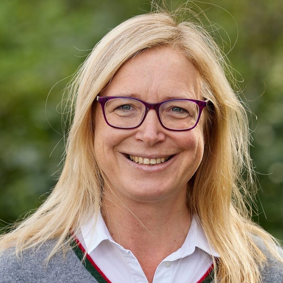
ruth birner-grünberger
MF2 Bioanalytics Coordinator
Technische universität Wien (TU Wien)
thilo hofmann
MF2 Mass Spec Coordinator
university of vienna

stephan krämer
MF2 Mass Spec Coordinator
university of vienna

laura liesinger
Technical Assistant MF2
Technische universität Wien (TU Wien)

federico lo nardo
Technical Assistant MF2
university of vienna

sarah reindl
PhD Student MF2
technische universität wien (tu wien)
MF3&4 Fluorescence In Situ Hybridization and Single-Cell Transcriptomics facility
Spatially resolved single-cell transcriptomics through parallel sequential FISH
Fluorescence in situ hybridization (FISH) with rRNA-targeted probes is a gold standard technique for the identification and visualization of microorganisms in environmental and clinical samples. This facility will make FISH, advanced confocal and super-resolution fluorescence microscopy, and digital image analysis techniques available to all CoE members. For selected projects, highly multiplexed FISH and multicolor imaging techniques for the parallel identification of multiple microbial species in complex samples will be adopted. For spatially structured microbiomes, 2D and 3D image analysis will extract important information, such as the spatial arrangement patterns of microbial populations.
Analyzing single-cell gene expression patterns within natural environments offers exciting opportunities to better understand microbial interactions and responses to perturbation. This facility will leverage signal amplification and multicolor imaging techniques to establish an mRNA-targeted FISH approach for spatially-resolved single-cell transcriptomics, which will be applied to detect selected gene transcripts directly in complex microbial communities.
holger daims
MF3&4 FISH Coordinator
university of vienna
michael wagner
MF3&4 Single-Cell Transcriptomics Coordinator
university of vienna
anton strunov
PostDoc MF3&4
UNIVERSITY of Vienna
stefan thiele
Technical Assistant MF3&4
university of vienna
Publications in Methods Facility 3+4
Rey Y. C., Kitzinger K., Lund M. B., Schramm A., Meyer R. L., Wagner M., Schlafer S. 2024, pH-FISH: coupled microscale analysis of microbial identity and acid-base metabolism in complex biofilm samples, Microbiome, 12(1), doi: 10.1186/s40168-024-01977-9
MF5 Stable Isotope Facility
Analysis of light elements in gases, solutes, and solids
The Stable Isotope Laboratory for Environmental Research (SILVER) provides high-end stable isotope ratio mass spectrometry (IRMS) techniques for light elements (H/D, 13C/12C, 15N/14N and 18O/16O) for bulk and compound-specific measurements.
The facility holds five IRMS coupled to the elemental analyzer, high-temperature pyrolysis unit, gas- and liquid-chromatography systems, and headspace gas preparation systems, as well as a high-resolution Orbitrap mass spectrometer with UPLC. SILVER specializes in (i) tracing isotopes into biomarkers (e.g., DNA, proteins, and lipids), (ii) isotope pool-dilution approaches to estimate gross biogeochemical process rates, (iii) measuring stable isotopes at their natural abundance, and (iv) fluxomics approaches based on isotope labeling. The services encompass advice on experimental approaches and evaluation of results, measurement execution, and help with the development of new methods.
Bernhard Lendl will complement the available techniques for stable isotope research by providing already available stable isotope selective mid-infrared laser gas sensors and novel portable devices in development.
bernhard lendl
MF5 Gas Sensors Coordinator
Technische universität Wien (TU Wien)
andreas richter
MF5 SILVER Coordinator
university of vienna
wolfgang wanek
MF5 SILVER Coordinator
university of vienna

erika kristel salas hernandez
PostDoc MF5
university of vienna

judith prommer
Technical Assistant MF5
university of vienna
MF6 Chemical Imaging Facility
Super-fast functional analyses of microbiomes
The CoE projects will benefit from a well-established NanoSIMS facility (unique in Austria), several spontaneous Raman micro spectrometry instruments coupled to fluorescence microscopes, and cutting-edge nano-infrared spectroscopy.
Furthermore, the projects will have access to a unique microfluidic-based Raman sorting device that enables us to sort thousands of microbial cells based on their isotopic composition or other chemical features. In addition, high-throughput single-cell isotope imaging in microbiome samples will be possible using our custom-made Stimulated Raman Spectroscopy (SRS) instrument, which is unique in Europe. It combines SRS with two-photon microscopy and enables, for example, quantification of the deuterium content (after heavy water incubation) of FISH-identified microbial cells at unprecedented speed.
The facility will push the limits of nano-IR spectroscopy by developing a dual-frequency comb spectroscopy approach for high-throughput chemical imaging.

bernhard lendl
MF6 Coordinator
technische universität wien (tu wien)

michael wagner
MF6 Coordinator
university of vienna
stefanie imminger
Technical Assistant MF6
UNIVERSITY of Vienna
margaux petay
PostDoc MF6
technische universität Wien (tu wien)
MF7 Microfluidics
and Lab/Organ-on-a-Chip Facility
Development of microscale assays for cell culture and analysis
This method facility offers analytical and development of microscale assays. We offer single-cell mass measurements in high throughput using a suspended microfluidic resonator system. The facility further contributes rapid prototyping technologies, microfluidics, CFD simulations, and optical and electrical biosensing systems, as well as bio-functionalization strategies to develop (portable) multifunctional lab-on-a-chip systems for cell culture and analysis.
Following the global trend of miniaturization, automation, and integration, the Lab-on-a-Chip laboratories maintain a range of rapid prototyping technologies and micromachining tools to develop computer-controlled microanalysis platforms, biochips, biosensors, micropumps, and micro-degassers. The facility is equipped to fabricate devices based on micromachining, lithography, soft- and hard polymer replication techniques such as casting, hot embossing, and micro-injection molding, as well as various 3D printing techniques for the fabrication of microdevices and microfluidic components facilitating controlled cell-to-cell interactions between roots, fungal hyphae, and bacteria.
peter ertl
MF7 Cell Chip Coordinator
Technische universität Wien (TU Wien)
martin polz
MF7 SMR Coordinator
university of vienna
alexandra lorenz
PhD student MF7
Technische universität Wien (TU Wien)

helena thumfart
PhD Student MF7
Technische universität Wien (TU Wien)
MF8 Protein Characterization Facility
Joint forces for structural biology of proteins
Genes of particular interest revealed by the CoE microbiome projects will be heterologously expressed (or native proteins and complexes purified directly from bacterial cultures), and their structures will be analyzed using integrative structural biology approaches.
The Djinovic lab at the University of Vienna has access to a blend of complementary molecular biophysics and structural biology techniques and is equipped for protein expression and purification.
The Sazanov lab at ISTA has access to state-of-the-art cryo-EM facilities, including a high-end 300 kV TEM Titan Krios for collecting high-resolution datasets, a 200 kV TEM Glacios for optimizing cryo-EM grids and preliminary data collection, and cryo-FIB/SEM Aquilos for preparing samples for cryogenic electron tomography.
Selected projects will be supported with enzyme discovery, profiling, and in-silico predictions, e.g., on secreted proteins by microbiome members using deep learning models trained on the sequence and structural features.
kristina djinovic-carugo
MF8 Coordinator
university of vienna
leonid sazanov
MF8 Coordinator
institute of science and technology Austria
jakob andersson
PostDoc MF8
institute of science and technology Austria
Foto: Clemens Fabry
georg mlynek
PostDoc MF8
university of vienna
MF9 Atomic Force Microscopy Facility
Resolving the ultrastructure of interactions with lasers
The atomic force microscopy (AFM) facility is equipped with four conventional AFMs, one fast-scanning AFM, one high-speed AFM, and one correlative AFM/Fluorescence Microscopy device, providing a variety of measurement modes.
AFM can perform topographical and mechanical imaging in physiological environments, resolving ultrastructure, mechanics, molecular, and cellular interactions at spatial dimensions from 1 nm to 100 µm with minute time resolution. High-speed Bio-AFM (HS-AFM) allows for filming molecular dynamics, conformational changes, and interactions of molecules in physiological environments with sub-nm spatial and 50 ms time resolution. Molecular Recognition Force Spectroscopy (MRFS) studies molecular interactions at the single-molecule level to determine interaction forces and energies. Cellular Recognition Force Spectroscopy (CRFS) deciphers interactions between microbes and hosts/biotic/abiotic surfaces on the cellular and molecular level. Topography and RECognition (TREC) Imaging localizes and distributes receptor binding sites on cells with nanometer resolution. Finally, Fluorescence Microscopy assists AFM in a combined AFM/Fluorescence microscope.
peter hinterdorfer
MF9 Coordinator
johannes kepler university linz
yoo jin oh
PostDoc MF9
johannes kepler university linz
MF10 Life Science Compute Cluster
High-performance computing infrastructure for computational life science
The Life Science Compute Cluster (LiSC) consists of data storage and high-performance computing facilities, and a user helpdesk. Parallel file systems with a total capacity of 1 PB are used for high-performance storage of original and derived data. Backups are performed on separate storage in a different location. The high-performance computing nodes provide >3,000 CPU cores and 30 GPUs and are used via a job scheduling system.
To facilitate easy and reproducible access to all relevant state-of-the-art scientific software, the LiSC provides a helpdesk to its users. The helpdesk not only supports users in case of problems with scientific computing, statistics, and data analysis but also takes installation requests for software versions.
The LiSC is connected to other high-performance computing facilities, namely the Vienna Scientific Cluster, allowing users straightforward access to additional computing power if needed.
thomas rattei
MF10 Coordinator
university of vienna
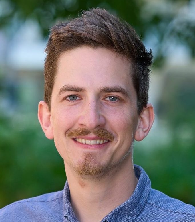
Foto: Clemens Fabry
lukas weilguny
PostDoc MF10
university of vienna

N.N.
Technical Assistant MF10
university of vienna


